Difference between revisions of "CT x-ray scanner"
From Noah.org
Jump to navigationJump to search| Line 22: | Line 22: | ||
|- | |- | ||
| [[image:section_4_3.png]] || [[image:section_4_2.png]] || [[image:section_4_1.png]] | | [[image:section_4_3.png]] || [[image:section_4_2.png]] || [[image:section_4_1.png]] | ||
| − | | | + | |} |
| − | + | ||
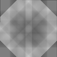
| − | + | Add (overlay) the four 1D radial sections together to get the CAT image. | |
| − | + | [[image:section_4_0.png]]+[[image:section_4_3.png]]+[[image:section_4_2.png]]+[[image:section_4_1.png]]=[[image:target_4_out.png]] | |
| − | image | ||
| − | |||
| − | |||
Revision as of 09:05, 13 December 2007
CAT Scanning
Here are some sample images that illustrate the process. The algorithm is quite simple.
In a real CAT Scan system the 1 dimensional slices would be taken from the horizontal row of a series of x-rays. In this demo I don't yet have the x-rays to work with so I synthesize the 1D bands from the target image that I want to regenerate. So given a target image I generate a series of 1D radial slices by rotating the target image and then averaging all values in the rows of the image. Then I rotate the slice back to the original angle.
Slices are synthesized from a 180 degree rotation of the target image.
The target and four 1-dimensional sections. The position of the sections corresponds to the angle of projection. |
 |

|
 |
 |

|
Add (overlay) the four 1D radial sections together to get the CAT image.
+
+
+
=